We use cookies to help you navigate efficiently and perform certain functions. You will find detailed information about all cookies under each consent category below.
The cookies that are categorized as "Necessary" are stored on your browser as they are essential for enabling the basic functionalities of the site. ...
Necessary cookies are required to enable the basic features of this site, such as providing secure log-in or adjusting your consent preferences. These cookies do not store any personally identifiable data.
Functional cookies help perform certain functionalities like sharing the content of the website on social media platforms, collecting feedback, and other third-party features.
Analytical cookies are used to understand how visitors interact with the website. These cookies help provide information on metrics such as the number of visitors, bounce rate, traffic source, etc.
Performance cookies are used to understand and analyze the key performance indexes of the website which helps in delivering a better user experience for the visitors.
Advertisement cookies are used to provide visitors with customized advertisements based on the pages you visited previously and to analyze the effectiveness of the ad campaigns.
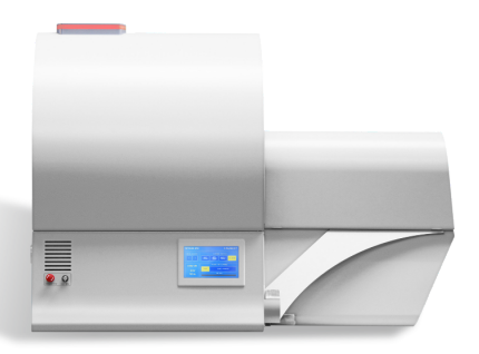
The large image field of view (up to 80 mm x 200 mm) allows full body mouse scanning with a single camera field of view. Variable X-Ray energy combined with a range of filters ensures optimal image quality for diverse research applications including pneumology, cardiology and body composition analysis. The system can perform scanning with continuous gantry rotation and in step-and-shoot mode with scanning cycles down to 7.2 sec.
Furthermore, the SKYSCAN 1278 in vivo micro-CT administers a low radiation dose to the animals allowing multiple scans in longitudinal preclinical studies without the risk of unwanted radiation-induced side effects. The fully integrated physiological monitoring package allows monitoring and controlling the animal’s wellbeing at all times through a video stream, ECG, temperature and breathing detection.
The SKYSCAN 1276 is complemented by 3D.SUITE. This extensive software suite covers GPU-accelerated reconstruction, 2D/ 3D morphological analysis, as well as surface and volume rendering visualization.
| Feature | Benefit |
| Integrated temperature support and physiological monitoring | Ensures animal well-being throughout the imaging session, and consistency between animals within a study. |
| Integrated inhaled anesthesia | Ensures ease of animal handling and consistent plane of anesthesia gas |
| 80 x 200 mm Field of View | Allows full body mouse and rat scanning (80 mm in diameter, 80 mm in length in a single scan) |
| Variable Magnification | Allows scanning bone and tissue samples with high spatial resolution down to 2.8 µm pixel size |
| 3D.SUITE Software | This extensive software suite covers GPU-accelerated reconstruction, 2D/ 3D morphological analysis, as well as surface and volume rendering visualization. |
| On-Screen Real-Time Dose Meter | The SkyScan1276 control software includes a real-time on-screen dose meter. It indicates an estimation of the dose absorbed by the animal body during scanning. The dose meter shows accumulated dose or dose rate. |
| X-ray Source | 20-65 kV, 50 W, with automated 4-position filter changer |
| X-ray Detector | 3 Megapixel active-pixel CMOS flat-panel, 1944 x 1536 pixels |
| Spatial Resolution | 75 µm pixel size 50 µm smallest pixel size up to 1536 x 1536 x 1566 pixels |
| Scanning Space | 80 mm in diameter 200 mm in length |
| Power Supply | 100-240V AC, 50-60Hz, 3A max. |
| Radiation Safety | < 1 µSv/h at any point on the instrument surface |
| System Dimensions | W x D x H: 85,0 cm x 124,0 cm x 96,0 cm Depth with open door: 162,0 cm Weight: 240 kg |
The 3D.SUITE Software – A perfect match for SKYSCAN 1276 CMOS Edition
Intuitive, simple, yet powerful – the 3D.SUITE software that comes with every SKYSCAN 1276 CMOS EDITION is designed to inspire finding out what’s inside. With the help of Genius Mode, even a novice user can intuitively start scanning right away. It helps optimize the scan conditions by choosing the appropriate filter and X-ray energy to achieve optimal image contrast, and by selecting the optimum exposure time and rotation step for efficient scanning.
The SKYSCAN 1276 CMOS EDITION is supplied with a GLP module, which allows administration of user rights in 3 levels and implementation of the necessary data protection according to GLP requirements.
NRECON | Reconstruction Software
The acquired 2D projection images are transformed into 3D volumes by the reconstruction software NRECON. Typical CT artefacts, such as beam hardening, ring artefacts and misalignment, are easily corrected. The SKYSCAN 1276 CMOS EDITION is supplied with GPU accelerated reconstruction providing results up to 10 times faster than traditional CPU-based reconstruction. GPU acceleration supports both conventional round CT and helical scanning.
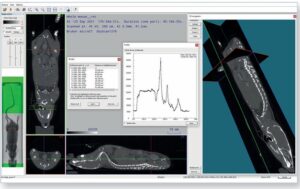
DATAVIEWER | Slice-by-slice Inspection
DATAVIEWER allows inspection of the reconstructed 3D volume using orthogonal slices in any direction. Objects can be rotated, repositioned, and resliced using their new orientation for more convenient visualization and saving of more efficient subvolumes. The software includes intuitive tools for measurement of 3D distances. 2D and 3D image registration enables the exact alignment of multiple scans of the same sample, acquired at different times.
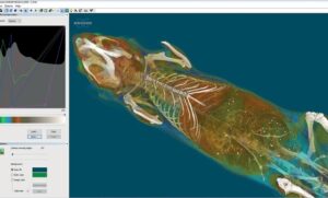
CTVOX | Realistic Visualization by Volume Rendering
CTVOX is an easy-to-use volume rendering package that provides precise control of visualization parameters, ensuring a realistic representation of all types of samples. CTVOX offers intuitive manipulation of the point-of-view, virtual slicing through objects, and full control of light, shadow, and surface properties. Creating attractive cover images and impressive movies has never been this easy.
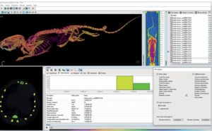
CTAN | 2D & 3D Image Analysis
Built over two decades based on direct feedback from scientists all over the world, CTAN is one of the most frequently used programs for quantitative image analysis. This package includes an extensive number of tools for region-of-interest selection, image segmentation and 3D measurements. Using the comprehensive library of embedded plugins or user-customized protocols, quantifying complex microstructures such as porosity, thickness, orientation, and many other properties is an easy task. Large sets of objects can simply be studied by automated batch analysis.
CTVOL | Built-in Surface Rendering
Surface models can be visualized in CTVOL, a flexible 3D viewing environment. Volumes can be exported in STL format, to allow 3D printing of the acquired scan data or further use in CAD and modelling programs.
Spine Microstructure
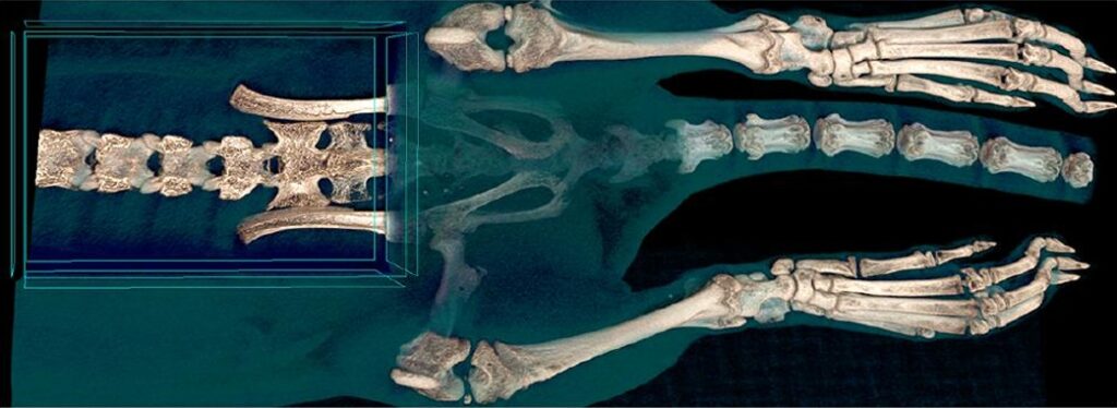
Volume rendering with virtual opening of the spine of a rat. Scanning protocol: 65 kV, “low dose” filter, 50 µm isotropic pixel size.
Full Body Mouse Scan
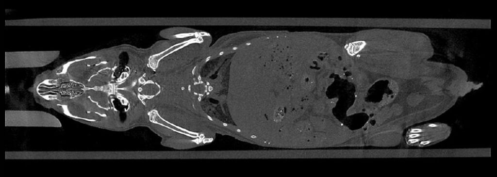
A reconstructed coronal virtual slice through a mouse body. No contrast agent. Scanning protocol: 65 kV, Al 1mm filter, 50 µm isotropic pixel size.
Time-resolved Heart Beat
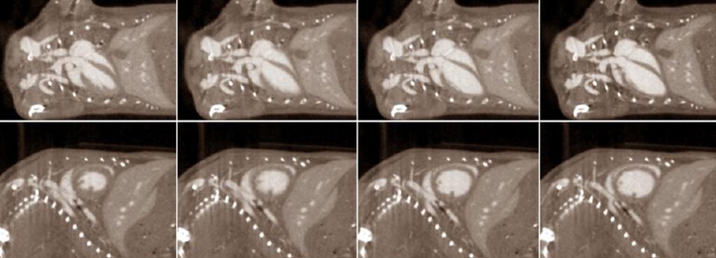
Coronal (top) and sagittal (bottom) reconstructed slices from four phases of the cardiac cycle. 4D scanning using contrast agent with data sorting according to ECG time marks.
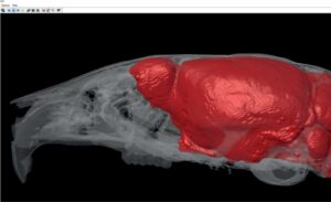
Fat Distribution Analysis

Volume rendering of the distribution of fat shown in orange inside the mouse body. No contrast agent. Scanning protocol: 60 kV, Al 0.5 mm filter, 50 µm isotropic pixels.
Using Contrast Agent
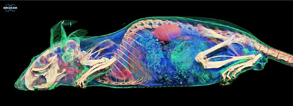
Volume rendering from a full body mouse scan with contrast agent injection. Scanning protocol: 65 kV, “low dose” filter, 50 µm isotropic pixel size.