
As image-guided radiation therapy (IGRT) has become the clinical standard, preclinical research labs require technology capable of replicating this method in mice and small animals. In modern labs, biology-guided irradiation is essential in translational radiation oncology studies to evaluate novel cancer treatments before clinical application.

The success of preclinical IGRT studies depends on three critical factors: Accurate 3D imaging to localize target areas, reliable treatment planning, and precise dose delivery.

How Innovation removes Barriers in Translational Radiation Oncology Research

XCELL TomoRad: The new, smart approach to preclinical IGRT. X-ray Irradiation / Image Guided Radiation Therapy for Mice and Rodents
The new XCELL TomoRad by KUBTEC solves common limitations in preclinical IGRT.

Until recently, there used to be a massive trade-off between affordability and precision among preclincal irradiation systems. Conventional cabinet X-ray irradiators provide an economical solution for broad-field irradiation, but they lack anatomical and functional guidance for precise targeting. And while existing preclinical IGRT systems offer high biology-guided accuracy, they require significant capital investment and highly specialized training – and workflows are often tedious.

But KUBTEC’s latest irradiation technology is bridging this gap, offering both high accuracy and a simplified workflow while making IGRT accessible to more labs:

The new XCELL TomoRad is a novel class of preclinical IGRT system, combining highly precise irradiation technology, advanced treatment planning, fast 3D imaging, and user-friendliness. This innovative solution enables scientists to perform image-guided preclinical irradiation more efficiently than ever before.

Limitations of Conventional Treatment Planning

Successful treatment begins with accurate planning, which requires precise 3D imaging.
Traditionally, micro-computed tomography (micro-CT or µ-CT) has served as the reference imaging modality for preclinical IGRT as it provides excellent anatomical detail in three dimensions.

However, full rotational µ-CT imaging requires hundreds of projection images, resulting in long scan times, large datasets, complex reconstruction, and additional radiation exposure. These drawbacks can be limiting factors in longitudinal studies where animals undergo repeated imaging and treatments.

Advanced Treatment Planning is now faster and easier

XCELL TomoRad solves these challenges by utilizing 3D X-ray tomosynthesis imaging.

Instead of acquiring projections through a full 360° rotation, X-ray tomosynthesis reconstructs volumetric anatomy from a limited-angle acquisition, which substantially reduces acquisition time by over 50% and requires fewer projection images for a full anatomical 3D reconstruction – resulting in a significantly lower radiation dose during imaging compared to conventional µ-CT protocols.

Moreover, image datasets become easier to manage, and treatment planning becomes more efficient. For most preclinical IGRT studies, 3D tomosynthesis provides sufficient anatomical detail to ensure accurate treatment localization, offering a smarter, faster alternative without the computational overload and time-consuming workflows of µ-CT reconstruction.

For tumor biology studies, the optional Bioluminescence Imaging (BLI) module can be used for functional imaging in addition to anatomical 3D tomosynthesis data, allowing precise targeting based on both structural and biological information.


Reproducible Animal Positioning for Precise Radiation Delivery

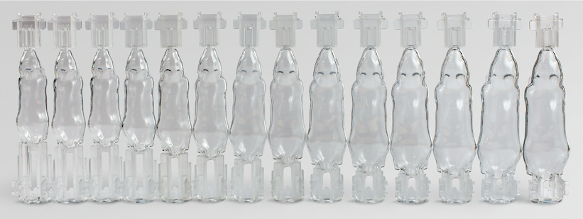
Body Conforming Animal Mold (BCAM) for Mice

Accurate imaging alone does not guarantee successful irradiation. Consistent animal positioning is equally critical for reproducible results and precise targeting.

The standard solution in IGRT irradiators for small animals is to rotate the beam source around a stationary animal. While effective, these systems rely on precise mechanical alignment between imaging and treatment components.

The XCELL TomoRad employs an alternative approach by maintaining a fixed X-ray source while rotating the immobilized animal in a geometrically fixed position.

At the center of this design is the Body Conforming Animal Mold (BCAM) — a reusable clamshell positioning system available in multiple sizes (in 1g increments) to accommodate different mice, always ensuring a perfect fit. The BCAM securely holds each animal  in a geometrically fixed position throughout imaging and treatment, ensuring highly reproducible results while minimizing motion that could compromise targeting accuracy.


Since the BCAM was originally designed for Bioluminescence Imaging (read our article on invivo Analytics here) and is made from a clear material with exceptional optical transmission, it can be used for BLI as well – and beyond the BCAM alone, there is also a full software solution for multimodal coregistered imaging with third-party systems, serving as a transport platform between preclinical imaging modalities.

Ultra-Fast Treatment: Higher Throughput, Shorter Anesthesia Time

Laboratories processing large animal cohorts must balance precision with efficiency. Reducing imaging and treatment time not only increases experimental throughput, but also supports animal welfare by minimizing anesthesia duration.

To accelerate radiation dose delivery, XCELL TomoRad incorporates the innovative Mag Mode feature (patent pending), which automatically positions the animal close to the X-ray source for dose rates of up to 16.25 Gy/min.

For lower dose rates, XCELL TomoRad offers flexibility for most preclinical applications, with a dynamic tube voltage range of 30–225 kV and up to 30 mA tube current.

Throughout imaging and irradiation, an integrated environmental chamber gives the operator full control over environmental conditions. Maintaining physiological stability throughout the procedure supports both experimental reproducibility and consistent 3R compliance for ethical animal research – and it helps maintain a suitable environment during cell irradiation studies.

Monte Carlo Algorithm for Accurate Dose Calculation

Hardware represents only one component of effective preclinical IGRT, but accurate treatment planning software is equally essential for ensuring consistent results and successful treatment. XCELL TomoRad utilizes comes with advanced treatment planning software by SMART Scientific, which incorporates Monte Carlo dose calculation algorithms and an user-friendly interface.

Monte Carlo simulation is widely regarded as the reference standard for modeling photon transport because it accurately predicts radiation interactions within heterogeneous tissues. This enables reliable dose estimation across complex tissue geometry in translational radiobiology research.


Bridging the Gap Between Cabinet Irradiators and Preclinical IGRT

Published research consistently demonstrates that accurate imaging, reproducible immobilization, and sophisticated dose calculation are fundamental to obtain reliable translational data. By combining three-dimensional tomosynthesis, stable animal immobilization, Monte Carlo-based treatment planning, and an efficient irradiation workflow, the XCELL TomoRad IGRT Irradiator bridges the gap between conventional cabinet irradiators and overly complex IGRT platforms for small animal research.

Rather than attempting to replicate every single feature of clinical IGRT systems, XCELL TomoRad focuses on the capabilities that really matter in practice for routine preclinical image-guided irradiation, and on making this technology more economically accessible.