
The X4 POSEIDON represents a paradigm shift in micro-computed tomography, merging the high-end capabilities of industrial-grade systems with the accessibility required for biomedical research.
Built on the legacy of the SKYSCAN series, this platform is designed to be the centerpiece of any life science lab, offering a non-destructive window into the microscopic 3D world of biological specimens.
Whether you are characterizing the porosity of a new pharmaceutical tablet, analyzing the mineral density of bone scaffolds, or tracking the development of a zebrafish model, the X4 POSEIDON provides the clarity needed for breakthrough insights. Its modular nature means the system is never obsolete; users can start with a basic configuration and add high-resolution detectors or transmission sources as their research focus evolves.
The X4 Poseidon is complemented by 3D.SUITE. This extensive software suite covers GPU-accelerated reconstruction, 2D/ 3D morphological analysis, as well as surface and volume rendering visualization.
The X4 POSEIDON is packed with innovative features that optimize the imaging workflow:
Multi-Vision Imaging
By incorporating two distinct detectors, the system can handle a massive variety of sample types. The 16MP sCMOS detector is optimized for high-contrast, high-resolution imaging with 2 µm focal spot, while the 7MP Flat Panel detector provides the speed and field-of-view required for larger specimens. You can also choose one detector and upgrade to Multi-Vision later!
GEM Plus™ Geometry
This proprietary technology allows the detector and sample to move independently. By optimizing cone beam geometry, GEM Plus™ significantly increases scan speed without compromising magnification ratio.
Integrated Automation
The front-mounted 15-position sample changer allows you to add or remove samples without interrupting active scans – enabling continuous, efficient high-throughput workflows.
Advanced X-ray Source Options
To meet different budgets and needs, X4 POSEIDON offers two sealed, maintenance-free X-ray sources.
The Reflection source provides high power for fast results on dense samples, while the Transmission source achieves a focal spot size of less than 2 µm. The sealed tubes require no external water cooling and offer extremely low cost of ownership.
In Situ Dynamics & 4D-CT
Go beyond static imaging with Bruker’s proprietary stages: Observe how your samples react to environmental stress in real-time using stages for heating, cooling, and mechanical testing (compression/tension), adding a 4th dimension (time) to your 3D data.
Comprehensive 3DxSUITE Software
The system is powered by the 3DxSUITE, a unified software environment that handles everything from acquisition to quantitative analysis. The interface is multi-language and designed for “walk-up” use by non-experts. For more details, see “Softwares” tab.
| X-ray Source | Reflection: Transmission: |
| X-ray Detector | Detector 1: Detector 2: |
| Nominal Resolution | Down to 2 µm (Transmission) Down to 5 µm (Reflection) |
| Max. Sample Size | 110 mm in diameter 300 mm in height |
| Sample Changer | 15 positions (up to 50 mm dia. / 80 mm height each) |
| Power Supply | 100-240V AC, 50-60Hz (Standard wall outlet) |
| System Dimensions | W x D x H: 125,0 cm x 60,4 cm x 69,0 cm Weight: 350 kg |
The 3D.SUITE Software – A perfect match for SKYSCAN 1276 CMOS Edition
Intuitive, simple, yet powerful – the 3D.SUITE software that comes with every SKYSCAN 1276 CMOS EDITION is designed to inspire finding out what’s inside. With the help of Genius Mode, even a novice user can intuitively start scanning right away. It helps optimize the scan conditions by choosing the appropriate filter and X-ray energy to achieve optimal image contrast, and by selecting the optimum exposure time and rotation step for efficient scanning.
The SKYSCAN 1276 CMOS EDITION is supplied with a GLP module, which allows administration of user rights in 3 levels and implementation of the necessary data protection according to GLP requirements.
NRECON | Reconstruction Software
The acquired 2D projection images are transformed into 3D volumes by the reconstruction software NRECON. Typical CT artefacts, such as beam hardening, ring artefacts and misalignment, are easily corrected. The SKYSCAN 1276 CMOS EDITION is supplied with GPU accelerated reconstruction providing results up to 10 times faster than traditional CPU-based reconstruction. GPU acceleration supports both conventional round CT and helical scanning.
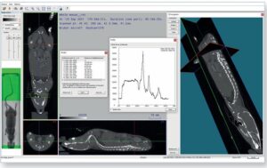
DATAVIEWER | Slice-by-slice Inspection
DATAVIEWER allows inspection of the reconstructed 3D volume using orthogonal slices in any direction. Objects can be rotated, repositioned, and resliced using their new orientation for more convenient visualization and saving of more efficient subvolumes. The software includes intuitive tools for measurement of 3D distances. 2D and 3D image registration enables the exact alignment of multiple scans of the same sample, acquired at different times.
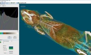
CTVOX | Realistic Visualization by Volume Rendering
CTVOX is an easy-to-use volume rendering package that provides precise control of visualization parameters, ensuring a realistic representation of all types of samples. CTVOX offers intuitive manipulation of the point-of-view, virtual slicing through objects, and full control of light, shadow, and surface properties. Creating attractive cover images and impressive movies has never been this easy.
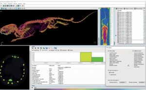
CTAN | 2D & 3D Image Analysis
Built over two decades based on direct feedback from scientists all over the world, CTAN is one of the most frequently used programs for quantitative image analysis. This package includes an extensive number of tools for region-of-interest selection, image segmentation and 3D measurements. Using the comprehensive library of embedded plugins or user-customized protocols, quantifying complex microstructures such as porosity, thickness, orientation, and many other properties is an easy task. Large sets of objects can simply be studied by automated batch analysis.
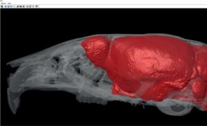
CTVOL | Built-in Surface Rendering
Surface models can be visualized in CTVOL, a flexible 3D viewing environment. Volumes can be exported in STL format, to allow 3D printing of the acquired scan data or further use in CAD and modelling programs.
Spine Microstructure
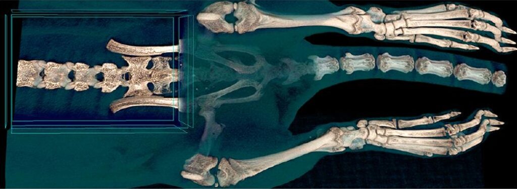
Volume rendering with virtual opening of the spine of a rat. Scanning protocol: 65 kV, “low dose” filter, 50 µm isotropic pixel size.
Full Body Mouse Scan
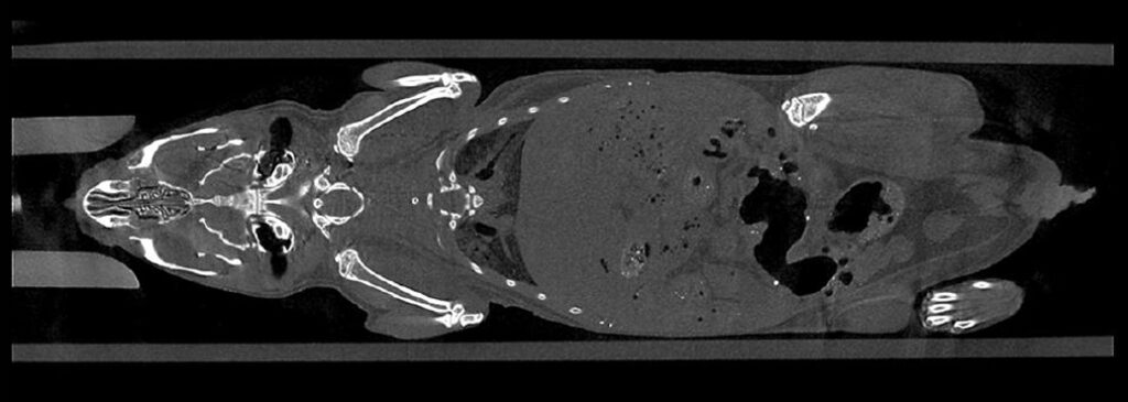
A reconstructed coronal virtual slice through a mouse body. No contrast agent. Scanning protocol: 65 kV, Al 1mm filter, 50 µm isotropic pixel size.
Time-resolved Heart Beat
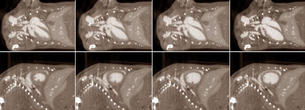
Coronal (top) and sagittal (bottom) reconstructed slices from four phases of the cardiac cycle. 4D scanning using contrast agent with data sorting according to ECG time marks.
Fat Distribution Analysis

Volume rendering of the distribution of fat shown in orange inside the mouse body. No contrast agent. Scanning protocol: 60 kV, Al 0.5 mm filter, 50 µm isotropic pixels.
Using Contrast Agent
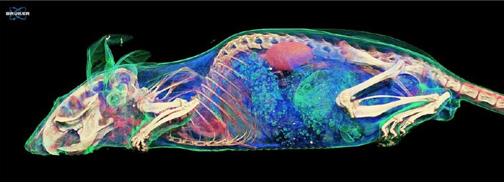
Volume rendering from a full body mouse scan with contrast agent injection. Scanning protocol: 65 kV, “low dose” filter, 50 µm isotropic pixel size.