
Beyond µ-CT: DXA for High-Throughput Bone Density Analysis in Mice

One of the primary challenges in preclinical skeletal research and metabolic phenotyping is balancing data depth and workflow efficiency.

While mapping bone microarchitecture may be necessary for some experiments, high-throughput longitudinal studies often rely on a simplified approach that eliminates tedious workflows and still yields valuable data: Bone Mineral Density (BMD) and Bone Mineral Content (BMC) analysis.

For years, micro-CT has been treated as a default method for bone assessments in the preclinical field, and for good reason – it provides an unsurpassed level of three-dimensional anatomical detail.

However, µ-CT analysis and 3D reconstruction require a substantial amount of time, knowledge and computational power per sample, and small animals are exposed to rather high doses of radiation, which needs to be taken into account, especially for repeated imaging.

But when research schedules demand faster workflows, larger cohorts, and safer protocols for consecutive imaging sessions, Dual-Energy X-ray Absorptiometry (DXA) has re-emerged as a tool of choice.


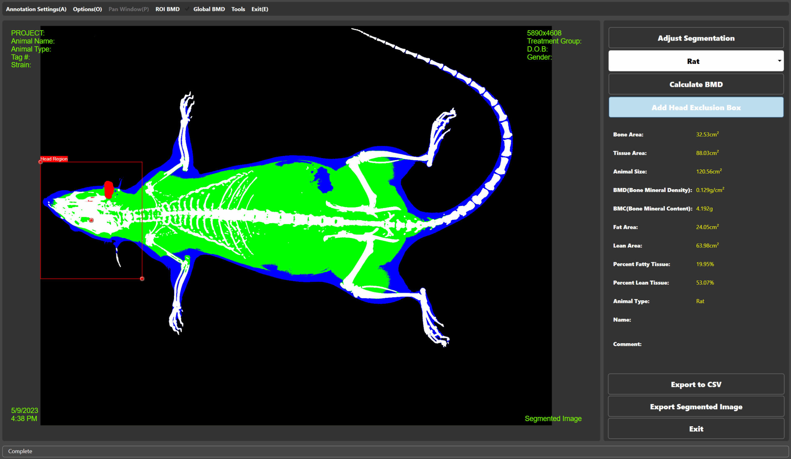
DXA Bone Mineral Density Analysis using KUBTEC Parameter X-ray imaging system
Bone Mineral Density Analysis using DXA
(using KUBTEC Parameter X-ray Imaging System + KUBTEC Digimus Software)

With modern low-dose, high-resolution X-ray imaging systems like KUBTEC Parameter or XPERT 80, DXA provides the ideal mix of precision, speed, low dose exposure and analytical ease for studies that focus on monitoring bone mineral density and body composition, rather than requiring detailed micro-CT images across multiple timepoints.

DXA acquisition and in-depth analysis are a matter of minutes, since DXA only requires a 2-dimensional X-ray image for reliable data.

For 3D anatomical imaging, 3D X-ray tomography provides a faster alternative to micro-CT, with only 90 seconds scan time and significantly lower radiation dose.


DXA vs. micro-CT: Benefits and limitations

DXA µ-CT

Primary Output


Areal Bone Marrow Density (BMD, g/cm²), BMC, Body Composition (Fat/Lean Mass). 

Lower spatial resolution based on 2D X-ray scan, but provides accurate data on body composition, tissue distribution and bone mineral density.

Volumetric BMD,
3D microarchitecture (BV/TV, Tb.Th)

Extremely high spatial resolution, ability to analyze and visualize bone microarchitecture

Scan Time
per Subject

< 10 – 30 seconds
5 – 15 minutes

Radiation Dose


Extremely low (< 1 mGy),
ideal for repeated scanning


High, repeated exposure can induce
radiation-driven bone changes

Data Management


Small file size per scan (megabytes), instant reports, easily handled by regular computers

Large files (gigabytes to terabytes), powerful computational infrastructure required for storage and processing

Data Analysis


Automated ROI selection possible, minutes per cohort


Complex 3D reconstruction, hours of manual or semi-automated 3D segmentation per mouse

KUBTEC X-ray Systems for DXA and BMD

When moving from theory to the lab bench, choosing the right instrument for your study goals is crucial. With over a decade of experience, KUBTEC Scientific has revolutionized preclinical DXA imaging. Their specialized systems are engineered to remove technical friction for preclinical scientists.

KUBTEC’s Parameter 2D and XPERT 80 systems feature ultra-high-resolution digital detectors to isolate the delicate skeletal anatomy of mice and rats. This allows for reliable evaluation of changes in bone density across tiny regions of interest with incredible reproducibility.

KUBTEC has a strong emphasis on ease of use:

  1. User-Friendly Interface, Easy Analysis:
    Place a subject in the cabinet, select the automated protocol, and obtain complete BMD, BMC, and body composition data in less than a minute.

  2. Seamless Software Automation:
    Define regions of interest (ROI) or select them automatically. The software separates fat, lean, and bone mass tissue – without the need for manual slice contouring.

  3. Safety Without Hassle:
    Fully shielded, self-contained X-ray cabinet – no need for specialized lead-lined rooms or additional safety equipment. 


Conclusion: Work Smarter, Scan Faster

If your lab is looking to explore mechanisms and advance treatments for osteoporosis, osteoarthritis, or metabolic syndrome, data throughput is critical. While micro-CT delivers complex 3D architecture, it introduces severe bottlenecks when used for large cohorts.

By integrating a KUBTEC X-ray imaging system into your experimental workflow, you gain access to rapid, low-dose, high-precision DXA capabilities. This allows you to scale up study sizes, track subtle bone and body composition changes over time, and focus your energy on new insights rather than processing heavy imaging datasets.