[Klose, A.; Paragas, N. (2018): Automated quantification of bioluminescence images. Nature Communications. DOI: 10.1038/s41467-018-06288-w]

Optical in-vivo imaging (OI), which includes bioluminescence imaging (BLI) and fluorescence imaging (FLI), is a well established and widely used modality in preclinical research. It’s a fast and easy imaging method that allows scientists to monitor luminescent signals in living small animals. The non-invasive nature of optical in-vivo imaging inherently aligns with the 3R’s (Replace, Reduce, Refine) of ethical animal science: it enables longitudinal tracking of biological processes in-vivo and eliminates the need to sacrifice cohorts at different timepoints.
However, optical in-vivo imaging has historically been limited by the need to manually draw ROI’s, low spatial and temporal resolution, and its inability to yield quantifiable biological data from the signal origin. It’s useful to quickly assess whether a change in signal intensity is observable, but OI data doesn’t provide much detail beyond that.
But invivo.ax has developed a new, advanced approach that overcomes these limitations, providing a wide range of new options far beyond traditional optical BLI, most importantly:
Reach out and secure your exclusive discount code for a free trial period. We’ll send you a demo bundle containing everything you need to get started.

To put it simply, optical in-vivo imaging systems are essentially highly sensitive photon counters. They offer a fast and easy way to assess if an effect is observable— for example, whether a signal can be measured or changes over time. OI is widely used for this purpose, but there are some limitations to keep in mind:
InVivo Analytics developed a new approach to overcome these limitations, and completely transforms the capabilities of optical in-vivo imaging. And the best news is, it does not require labs to purchase an entirely new in-vivo imaging unit – you can likely use the system you already have. While it works more accurately with a high-end system like the LAGO, multiple manufacturers are supported and support for new systems is continuously added.
By integrating innovative hardware and software, invivo.ax offers a complete platform that finally enables researchers to translate optical imaging surface signals to quantifiable data about biological processes right from the signal’s source.
invivo Analytics overcomes traditional limitations through three key innovations:
1. Body-Conforming Animal Mold (BCAM)
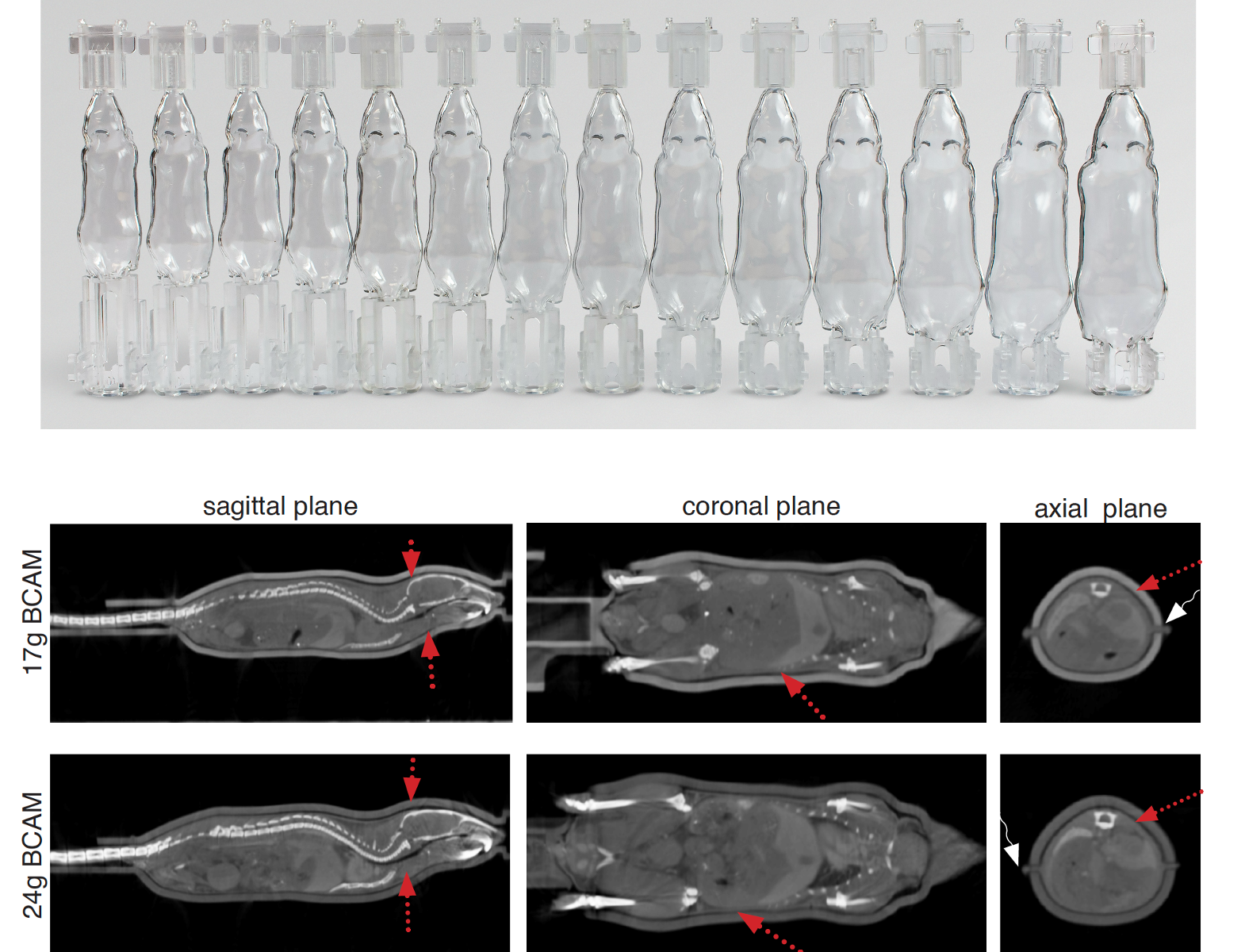
The BCAM is an optically transparent, rigid clam-shell shuttle scaled to an animal’s weight that holds mice in a fixed position. This physical constraint ensures consistent, standardized anatomical geometry across animals of varying sizes. BCAM’s are available in 1g increments, always ensuring excellent skin-to-BCAM contact and a snug fit. By standardizing the animal’s geometry, the BCAM drastically improves data congruency, even beyond optical imaging: it allows for easy coregistration with other imaging modalities (like µ-CT) across manufacturers, while still maintaining the animal’s position.
2. Statistical Mouse Atlas (OPM)
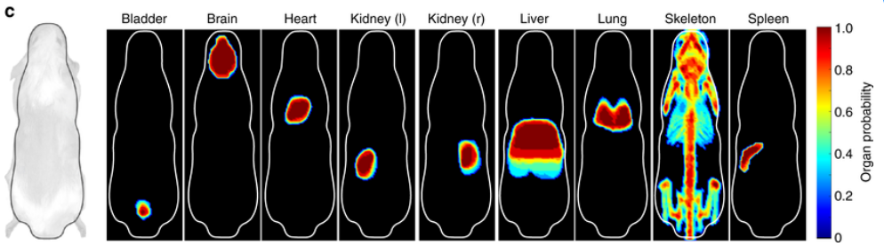
The Organ Probability Map (OPM) functions as a statistical mouse atlas built from segmented CT scans of multiple mice in the standardized geometrical position inside the BCAM. It provides an immediate, highly accurate anatomical reference. Because the OPM is intrinsically coaligned to the BCAM shape, it allows for automatic ROI drawing and analysis of target areas (like organs, body parts or tumors) across entire cohorts, completely eliminating the need for manual ROI drawing and operator bias (while still giving users the option to draw ROI manually if needed).
3. 3D Bioluminescence Reconstruction Software
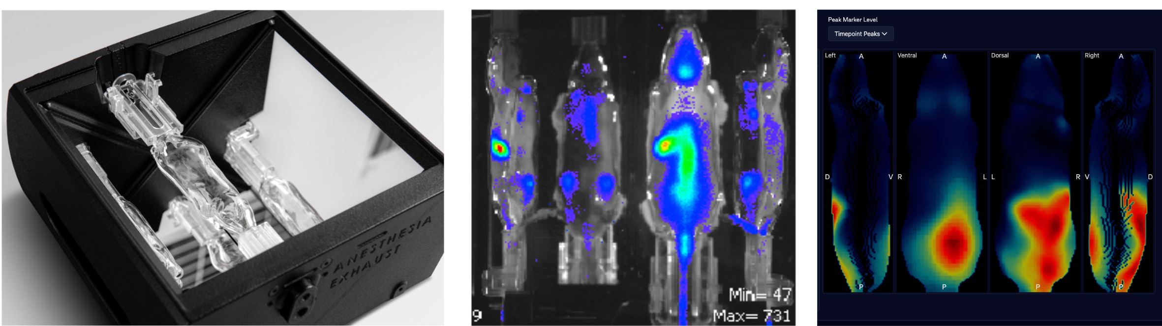
The cloud-based web app utilizes advanced reconstruction algorithms to convert 2D surface light measurements, obtained from four different angles using the invivoPLOT mirror gantry, into precise 3D maps of the photon emission density inside the tissue at source. It utilizes the Organ Probability Map to take into account which tissue the signal has travelled through and how the light got scattered and attenuated, allowing for reconstruction of signal intensity at source, and signal distribution in three dimensions.
The backbone of this technology is a mirror gantry, called invivoPLOT, that is used with a BCAM. The data is then uploaded to the cloud-based invivo.ax software, accessible as a web app. This software makes it incredibly easy and fast to reconstruct and analyze whole-body BLt data—often in under 60 seconds, even with a standard laptop, since the computational workload is managed by powerful servers via cloud. Because the BCAM provides a known surface geometry beforehand, the computationally heavy projection matrices are precalculated, allowing the software to translate 2D surface intensities into 3D bioluminescence images almost instantaneously.
Ultimately, researchers can linearly translate this reconstructed photon emission density into quantifiable units, such as a spatial map of bacterial colony-forming units (CFU/mm^3) with high fidelity and accuracy that matches or even surpasses the accuracy of other conventional methods, as shown in this validation study.
By achieving standardized positioning, operator-independent ROI analysis, and actual 3D source quantification using standard optical hardware, InVivo Analytics drastically increases data reproducibility. Higher reproducibility means researchers can achieve robust statistical power with smaller sample sizes, marking a major leap forward in adhering to the ethical 3R guidelines for animal research.